Sunday, 20 January 2013

Aribex ships handheld x-ray system


Aribex ships handheld x-ray system
By DrBicuspid Staff
July 17, 2008 -- Handheld x-ray developer Aribex has begun shipping its Nomad Pro digital x-ray system.
Introducing CEREC® Omnicam.The most perfect CAD/CAM camera ever
SironaSlim, elegant design for easier intraoral access, fast photorealistic color imaging, and powderless convenience make the new CEREC Omnicam the most precise, easy-to-use CAD/CAM
camera ever.
Nomad Pro includes a full-color LCD display screen, preset exposure settings, and additional time-saving features, according to the Orem, UT-based firm. It weighs 5.5 lb and is suitable for general-purpose dental use, Aribex said.
The system has received U.S. Food and Drug Administration clearance, Aribex said.

ADA and Congress face off over amalgam separators


ADA and Congress face off over amalgam separators
By Rabia Mughal, Contributing Editor
July 16, 2008 -- The ADA is standing its ground on amalgam separators. In hearings last week on Capitol Hill, an ADA representative reiterated the association's position that requiring all U.S. dentists to install amalgam separators in their offices would be an unnecessary financial burden.
But government officials and public advocacy groups believe that cost is not the real issue. They are accusing the ADA and other dental associations of deliberately blocking attempts to regulate the use of amalgam separators.
Testifying before the domestic policy subcommittee of the House Committee on Oversight and Government Reform at a hearing to assess state and local regulations to reduce dental mercury emissions, William J. Walsh, outside counsel for the ADA on amalgam wastewater issues, offered oral and written testimony supporting the ADA's efforts to create "voluntary, cooperative partnerships" to reduce the environmental impact of dental mercury emissions.
He noted that the ADA has issued several best management practices (BMPs) for handling waste amalgam. These BMPs include standard control methods, recycling of collected amalgam, and, since last fall, the use of amalgam separators.
Walsh also argued, however, that dentistry's contribution to surface water mercury is very minor.
"Dentistry contributes less than 1% of the total mercury found in our lakes and streams," Walsh said in a prepared statement.
Even though 40% to 50% of mercury that enters wastewater comes from dental offices nationwide, wastewater treatment plants catch most it, according to Walsh. Eventually, dental offices contribute only 0.4% of the mercury that goes into surface water, he said in an interview with DrBicuspid.com.
If every dental office nationwide installed an amalgam separator, it would have a minimal impact on this already small number, Walsh added added. Currently only nine states require dental offices to install amalgam separators.
"If you take a standard dental office without an amalgam separator, 99% of mercury is captured by the wastewater treatment plant, while those with a separator get 99.2% of their waste captured," Walsh said. "Eventually, it is almost the same amount. The only difference is that in offices with an amalgam separator, the mercury gets captured before it goes into the sewerage system."
A mandatory amalgam separator program would be an administrative burden and increase the cost of dental care, as well as the cost of regulating dentistry, he said.
The overall cost to a dentist to install an amalgam separator includes buying the separator, installing it, and annual operation and maintenance fees, Walsh explained. The capital cost and installation are approximately $1,200, and the annual cost of owning and operating an amalgam separator is around $770.
A voluntary program is more appropriate for attaining mercury reduction from a professional group like dentists than a "command-and-control" approach, he added.
"The ADA believes that the issue of installation should be decided on a case-by-case basis based on site-specific conditions," Walsh said.
The EPA also favors a voluntary program when it comes to small businesses, he added.
"If it is not effective, the government can always make it mandatory," Walsh toldDrBicuspid.com.
The three subcommittee members at the hearing were not receptive to the ADA's argument.
"I just have to say to the unbiased observers that it seems to me the ADA has a resistance to mandatory separators that is almost theological," said Rep. Dennis Kucinich (D-OH), who chairs the domestic policy subcommittee, according to an ADA news article. "Are there product liability concerns or a fear of class action in the offing? Is there something deeper here?"
Michael Bender, director of the Mercury Policy Project, a public advocacy group that supports mandatory amalgam separators, testified that separators are the most cost-effective method of controlling mercury pollution from dental offices.
The total cost of installing separators in all general practices across the U.S. would be around $116,999,141, Bender testified. Considering 60,000,000 amalgam fillings are placed each year, the separator cost would only be $1.95 per filling, he argued.
"Cost to remove mercury at a wastewater plant is $21 million per pound, or $46,000 per gram, compared with amalgam separator cost of $1.95 for an average mercury filling removal," he said.
He accused the ADA and state dental associations of blocking amalgam separator initiatives.
"The California Dental Association was the sole opponent of Assembly Bill 966 in 2005, authored by Assembly Member Lori Saldaña, and stopped the bill in the Assembly. The bill would have mandated separators," according to a report presented by the Mercury Policy Project at last week's congressional hearing. "Last year, the Pennsylvania Dental Association blocked a proposed ordinance by the Philadelphia City Council would have required most dentists residing in Philadelphia to install amalgam separators."
Asbjorn Jokstad, D.D.S., Ph.D., head of the department of prosthodontics at the University of Toronto, agrees that amalgam separators should be mandatory.
They are the only effective way of minimizing mercury release in the water supply as they capture the small particles, he stated in an e-mail to DrBicuspid.com. With other measures, larger amalgam particles are caught and their weight gets included in the overall percentage of mercury released into the environment, he explained.
"Although these larger particles are also of some environmental concern, they are of less worry since they will not undergo the mercury methylation process that occurs in aquatic milieu," he concluded.
"The record clearly shows that voluntary programs are not effective at convincing dentists to install amalgam separators," Bender stated in his testimony before Congress. "To prevent future pollution and costs, separators should be required and amalgam phased out."

Washington dental board too soft on death cases?


Washington dental board too soft on death cases?
By DrBicuspid Staff
July 16, 2008 -- A Seattle dentist who recently resigned from the Washington state dental board has questioned the consistency and thoroughness of its review process in three dental death cases, according to a recent seattlepi.com story.
In the past three years, at least three patients died after dental procedures and all three cases were closed without charges or a full hearing by the Washington State Dental Quality Assurance Commission, the story reported. In one case, the dentist/doctor who was excused by the dental board has been charged with unprofessional conduct by the state's medical board for the same surgery.
"Because no action was taken by the dental board in any of the cases, none of the deaths shows up on the state Web site where consumers can check their dentists' histories," reported seattlepi.com.
Other states, such as California and Texas, do a full investigation of all dentistry-related deaths, the story revealed. However, under Washington law, a death case can be closed before an investigation based on a dentist's summary of events.
The seattlepi.com story provides a detailed account of all three cases. To read the full story click here.

Study catches dentists dirty-handed


Study catches dentists dirty-handed
By Laird Harrison, Senior Editor
July 15, 2008 -- Do you wash your hands between patients? Do you use alcohol sanitizers each time you change gloves? Surprisingly, most dentists aren't following the hand hygiene recommendations of the Centers for Disease Control and Prevention (CDC), a study of New York practitioners found.
The study, published in the July issue of the Journal of the American Dental Association(Vol. 139:7, pp. 948-957), found that many dentists were not washing their hands frequently enough, and even more were not using alcohol-based hand sanitizers as often as recommended.
"It's surprising how many dentists don't clean their hands," commented Joan Duggan, M.D., a University of Toledo researcher who has done similar research in physicians. (The study's authors, most based at Columbia University, could not be reached for comment.) But she noted that medical doctors fail to comply with the recommendations in approximately equal numbers.
In what appears to be the first study of dentists' hand hygiene in the U.S., the researchers sent surveys to 352 general dentists who were members of the New York State Dental Association. They received 234 responses.
The survey distinguished between washing with soap and water and disinfecting with alcohol sanitizers. According to the CDC's "Guidelines for Hand Hygiene in Healthcare Settings -- 2002," disinfecting with alcohol sanitizers is more effective.
The study authors boiled down the CDC recommendations in this way:
  • Whenever your hands are visibly soiled, wash with soap and water for at least 15 seconds.
  • After removing your gloves, and before putting them on again, use an alcohol sanitizer.
  • If you are doing a procedure that penetrates a sterile surgical site, wash with soap and water and disinfect with an alcohol sanitizer before putting on your gloves.
Most dentists in the survey -- 71% -- said they washed their hands at the start of the day, 1% start with disinfecting and another 22% do both, leaving a benighted 6% who do neither one.
But 19% admitted that they neither wash nor disinfect their hands between patients. Another 24% said they don't clean their hands each time they remove their gloves. And -- in the clearest violation of the recommendations -- 65% didn't use alcohol sanitizers after removing gloves.
Shockingly, 20% apparently didn't wear gloves at all.
Why aren't dentists following the guidelines? Some may be confused by conflicting messages. The CDC's 2002 guidelines describe alcohol sanitizers as superior to soap-and-water washing, while the CDC's "Guidelines for Infection Control in Dental Health-Care Settings -- 2003" simply mention alcohol sanitizers as an alternative to soap and water.
Alcohol sanitizers truly are superior, according to Dr. Duggan. "I think the evidence is pretty damn good." (If you're shopping, you may want to visit the Environmental Protection Agency's list of products effective against various pathogens.)
Another problem may be that some dentists have too much confidence in their gloves. Although latex gloves appear impermeable, imperfections in them can admit microbes. The use of gloves only reduces the risk of contamination by 70% to 80%, according to the CDC. And gloves also encourage microorganisms to grow by creating a warm, moist environment.
Clearly, many dentists need to be reminded of such facts, Dr. Duggan said. "Despite the fact that the message has been out there for 150 years, it's only gradually getting better."

Copyright © 2008 DrBicuspid.com

Dental braces stop bullet, save life


Dental braces stop bullet, save life
By DrBicuspid Staff
July 14, 2008 -- A Michigan teenager who was critically injured in a recent gun fight appears to have been saved by his braces, according to an NBC news story.
Anthony Pittman, 18, was hit in the mouth with a .45-caliber bullet last week. The bullet split into pieces on contact with his braces, according to police.
"We believe what happened is that the bullet was split, in part by the braces, so it fragmented and continued into his mouth, tongue, and teeth," said Captain Wendy Keelty of the Pontiac Police Department in the NBC story. "But because it was fragmented, it didn't penetrate through the back of his neck."
Pittman has several broken teeth and a severely injured tongue. It will be a while before he is able to resume speech, according to his doctors.

Copyright © 2008 DrBicuspid.com

Oral piercings: Where fashion and dentistry clash


Oral piercings: Where fashion and dentistry clash
By Rabia Mughal, Contributing Editor
July 14, 2008 -- Being a teen's dentist is not easy. You are already battling problem trends such as crystal meth, smoking, questionable eating habits, and even diabetes.
Now a new study has pushed an old enemy to the forefront: oral piercings.
Researchers at the recent International Association for Dental Research (IADR) session in Toronto reported that the most common complications related to oral piercings are chipped, fractured, or cracked teeth and gingival recession. Less common but life-threatening complications include Ludwig's angina, Lemierre's syndrome, and hemorrhage.
“Health professionals should be aware and knowledgeable of these risks in order to provide their patients appropriate advice.”
The researchers noted that oral piercings are most popular among younger patients between the ages of 18 and 30, and the tongue and lip are the most common piercing sites. They conducted a systemic review of research on the adverse effects of oral piercings over the last 12 years.
"Piercers are usually unlicensed and untrained," the authors noted. "Health professionals should be aware and knowledgeable of these risks in order to provide their patients appropriate advice."
A previous study looked at research published between 1992 and 2007 and found that 10% of all New York teenagers have some kind of oral piercings, compared to about 20% in Israel and 3.4% in Finland (American Journal of Dentistry, December 2007, Vol. 20, pp. 340-344).
The authors reviewed articles on complications from oral piercings. From the reviewed literature, the lowest recorded rate of swelling and infection after a piercing by a study was 24%, while the highest rate noted was 98%. Pain or tenderness ranged between 14% and 71%, while speech interference came in at 14% to 51%. Numbness, eating difficulties, and bleeding were common short-term postoperation side effects as well.
Jewelry-induced complications included tooth fracture (13% to 41%), gingival recession (19% to 68%), and ingestion of jewelry (7% to 34%). Other side effects included taste interference and halitosis.
"There are short-term complications to piercings in low percentages of teens, and in rare cases a piercing to the oral cavity can cause death," stated Liran Levin, D.M.D., a clinical instructor at the Maurice and Gabriela Goldschleger School of Dental Medicine at Tel-Aviv University and an author of the 2007 study, in a recent press release. "Swelling and inflammation of the area can cause edema, which disturbs the respiratory tract."
"There is a repeated trauma to the area of the gum," he added. "You can see these young men and women playing with the piercing on their tongue or lip. This act prolongs the trauma to the mouth and in many cases is a precursor to anterior tooth loss."
Dan Peterson, D.D.S., who practices in Gering, NE, has had several teen patients come in with swelling and infection because of an oral piercing, and had to talk them into taking the piercing out and letting the area heal.
"I tell my patients that, even if they do not have any immediate infection, there will be some piercing-related complications in the future, so overall it's just a bad idea," Dr. Peterson said. "There are too many things that can go wrong."
With lip piercings, the jewelry constantly rubs against the gum tissue, which causes erosion over the long run, he said. In addition, biting down hard on a tongue piercing can chip teeth. People with tongue piercings are also subject to infections, and a severe tongue infection can cause breathing problems.
The piercing community does not deny these risks.
"There are always risks attached to oral piercings," said James Weber, president of the Association of Professional Piercers (APP). "We try not to downplay them."
Chipped teeth, gum recession, and swelling are potential side effects of piercing, he said. But appropriate placement and the right kind of jewelry can lower those risks significantly, he added.
"Right now, most piercing salons have to abide by very few regulations, if any, and that is why the clients need to take responsibility," Weber said.
The APP urges people to go to a piercer who is skilled, experienced, and uses sterile instruments, jewelry, and disposable needles. Clear aftercare instructions should also be provided. According to the APP, if a piercer tells their clients to treat a piercing with harsh soap, ointment, alcohol, or hydrogen peroxide, the studio is not keeping up with industry standards. The APP recommends sea salt and a mild liquid soap. (For more details on ensuring safer piercings, you can direct your patients to the APP Web site, which has information on choosing a piercer and oral piercing risks and safety measures.)
ADA consumer advisor Matthew Messina, D.D.S., who practices in Cleveland, has done fillings and crowns on chipped teeth and dealt with several infections -- all related to piercings.
An oral piercing is an open wound, he pointed out. Unlike an ear piercing, if you remove the jewelry from an oral piercing, the hole closes because it is moist tissue.
An antibacterial rinse should be a mandatory part of the daily routine for anyone with a tongue piercing, as should cleaning the piercing bar regularly to avoid tartar buildup, Dr. Messina said. He also recommended having patients seek immediate medical help if there is any redness, swelling, or pain around the piercing.
In the long run, however, the dental community's advice: Don't do it!
"Teenagers are not easy to manage," Dr. Levin noted. But "try where possible to dissuade your teen from getting a piercing," he said. "They will thank you when they are older."

Copyright © 2008 DrBicuspid.com

Beyond x-rays: Part III -- OCT brings early decay to light


Beyond x-rays: Part III -- OCT brings early decay to light
By Kathy Kincade, Editor in Chief
July 11, 2008 -- Imagine being able to see inside a tooth with 10 times the detail provided by the best x-ray system on the market. Imagine being able to do this in near real-time, chairside, with little more than the press of a button.
Introducing CEREC® Omnicam.The most perfect CAD/CAM camera ever
SironaSlim, elegant design for easier intraoral access, fast photorealistic color imaging, and powderless convenience make the new CEREC Omnicam the most precise, easy-to-use CAD/CAM
camera ever.
Now, how much would you be willing to pay for all that?
If your answer is around $25,000, you may be in luck. Next-generation laser-based technologies that improve early detection and diagnosis of caries and oral disease are nearing commercialization. While the cost of these devices may cause you to balk, the companies developing them believe their diagnostic advantages will convince you to reconsider.
Optical coherence tomography (OCT) currently leads the pack in next-generation diagnostic techniques being developed for dentistry. It is similar to ultrasound except that it uses light rather than sound waves. By measuring the time delay and magnitude of the reflected light, it creates micron-scale images (see video below). The imaging depths are typically up to 3 mm in hard tissue, which is shallow compared with ultrasound. However, OCT can provide much higher resolution images than ultrasound.
            Video courtesy of Dr. Craig Gimbel, Lantis Laser.
Having trouble viewing this clip?
Click here to download the free Flash player.

In fact, with resolution 10 times better than x-rays, the promise of OCT in dentistry is that it will allow for early detection of dental caries, microstructural defects, and periodontal disease in both 2D and 3D. OCT can image both hard and soft tissue, something that x-rays cannot do, and it can image decay in the pits and grooves of the biting surfaces, where 90% of decay begins. Detail unavailable with x-ray is obtainable with OCT, allowing for fast and accurate assessment of restoration quality.
OCT is already a well-established imaging modality in ophthalmology but has yet to find its footing in dentistry -- despite the fact that dental researchers have been experimenting with it since the late 1990s. In fact, the first OCT images of teeth were generated in 1998 (Optics Express, September 1998, Vol. 3:6, pp. 230-238).
Two years later, researchers from the University of Connecticut School of Dental Medicine and Lawrence Livermore National Laboratory, working with a custom-built OCT system, concluded that OCT has several advantages compared with conventional dental imaging. "OCT images exhibit microstructural detail that cannot be obtained with current imaging modalities [and] visual recordings of periodontal tissue contour, sucular depth, and connective tissue attachment now are possible. The internal aspects and marginal adaptation of porcelain and composite restorations can be visualized. This new imaging technology is safe, versatile, inexpensive, and readily adapted to a clinical dental environment," they wrote (Journal of the American Dental Association, April 2000, Vol. 131:4, pp. 511-514).
More recent studies have found that OCT shows promise for applications beyond caries detection. In a study on vertical root fractures in 25 mandibular premolars, researchers from the Erasmus University Medical Center in Amsterdam found that OCT is a promising nondestructive method of diagnosis for vertical root fractures. "We believe that a specially designed endodontic probe will improve visualization for conventional endodontic treatment and could lead to better decision making and elevated standard of care," they concluded (Journal of Endodontics, June 2008, Vol. 34:6, pp. 739-42).
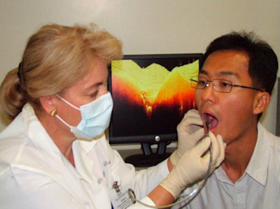
OCT captures cross-sectional images of hard and soft dental tissue up to 3 mm deep into the tissue. These images are then displayed individually in real-time on a chairside monitor and can be saved to the patient digital file. Image courtesy of Dr. Craig Gimbel, Lantis Laser.
Edwin Parks, D.M.D., M.S., director of dental radiology at Indiana University, agrees.
"I see OCT as a good way to detect cracks in teeth versus looking for caries -- and cracked teeth are a huge issue in dentistry," he said. "I would like to see it work for the benefit of the patient so you don't end up doing a $1,000 procedure on a tooth that is doomed. If you have a cracked tooth and it is cracked into the pulp, the outcome is probably not very good. And if you know this ahead of time, you probably won't dink around with doing a root canal and putting a crown on it."
OCT could play a role in orthodontics as well. A recent publication by Cosmin Sinescu et al (Proc. SPIE Int. Soc. Opt. Eng. 6991, 69911U, May 2008) showed how using OCT to examine the outcome of different bracket-bonding materials and procedures could affect stability of bracket retention and minimize failures.
"The biggest horror for an orthodontist is to remove the brackets on the anterior teeth and see white spot lesions buried underneath the bracket and the adherent bonding agent," said Craig Gimbel, D.D.S., former president of the Academy of Laser Dentistry. "OCT will enable you to image through the bracket [before it is removed] to determine what is going on underneath."
OCT also has soft-tissue imaging capabilities, such as determining stages of dysplasia in oral cancer and monitoring periodontal ligaments, Dr. Gimbel noted. In a recent study, researchers from the department of orthodontics at Ulsan University Hospital in South Korea observed structural variations of the periodontal ligament in the maxillary anterior teeth of white rats under different orthodontic forces through the use of OCT imaging (Medical & Biological Engineering & Computing, June 2008, Vol. 46:6, pp. 597-603).
“This may lead to a potential application of OCT for monitoring and determining maximum appliance load forces to prevent the tearing of the periodontal ligament in humans during orthodontic procedures,” Dr. Gimbel said.
Commercial development
Dr. Gimbel is also the clinical director for Lantis Laser of Denville, NJ, the only company so far commercializing OCT for the dental market. Lantis has an exclusive license to dental applications for OCT through agreements with Lawrence Livermore National Lab and another OCT developer, LightLab Imaging.
"Our OCT system will pick up the presence of decay much sooner [than other optical technologies] with much less decay present," said Stan Baron, president and CEO of Lantis Laser. "And it provides a cross-sectional image that correlates with with histology, indicating both how much decay there is and where it is located, not just an auditory signal that indicates that some degree of decay is present."
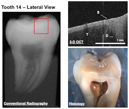
A conventional radiograph, OCT image, and histology of the same tooth [(T) tooth, (S) sealant, and (D) decay]. Note that no decay is seen in the radiograph, while both the OCT image and histology confirm the presence of decay. Photo courtesy of Petra Wilder-Smith, D.D.S., Ph.D.; Dr. Jae Ho Baek; Dr. Zhongping Chen; and Dr. Yehchen Ahn of the Beckman Laser Institute at the University of California Irvine.
Dr. Gimbel believes that OCT brings something to the dental community that it's never had: the ability to visually distinguish between cavitated and noncavitated lesions earlier than ever before.
"This is truly minimally invasive dentistry," he said. "If you are trying to determine whether a lesion is cavitated or noncavitated simply by looking only at the surface, you don't know completely. But OCT allows you to see the microstructural changes such as demineralization early on in the development of the decay. X-rays are a gross image, while OCT is a microstructural image."
This distinction has potentially important implications for a number of dental procedures, Dr. Gimbel added. For example, recent research has shown that it is acceptable to place a sealant in a noncavitated area of decay because the bacteria will die under the composite. But how do you know if the lesion is cavitated or noncavitated to begin with?
"This is what OCT can do," Dr. Gimbel said. "It provides the missing puzzle piece by enabling you to monitor the lesion underneath the composite. There is no way to do this with an x-ray if the lesion is noncavitated."
Lantis Laser has spent the last few years developing a time-domain OCT system for dentistry and had expected to introduce the product this year at a price of $20,000 to $25,000. But in March the company announced that it is now is partnering with Axsun Technologies to upgrade to a faster and more sensitive OCT technology. As a result, Lantis says commercial release of its first OCT system will be delayed until next year. But the trade-off is worth it, according to Baron.
In the long run, however, the price may be a sticking point. According to Greg Smolka, author of the market research report, "Optical Coherence Tomography: Technology, Markets, and Applications, 2008-2012," Lantis is hoping to achieve 5% market penetration in four years after introducing its OCT system. Digital radiography, he said, has taken 10 years to achieve a 20% market penetration in dentistry, despite the technology being very similar to traditional x-ray.
"OCT is a promising technology that can offer new and perhaps better information to the dentist, and there is certainly a large market for such an advanced diagnostic tool, assuming strong clinical efficacy is proven," Smolka said. "However, for OCT systems to reach widespread acceptance in the dental office, they must prove they are worth the expensive price tag by offering significant diagnostic advantages over traditional and other emerging imaging techniques."

Most-effective dental braces are the least attractive, survey finds


Most-effective dental braces are the least attractive, survey finds
By DrBicuspid Staff
July 10, 2008 -- When it comes to the attractiveness of orthodontic braces, less metal is better, according to a survey published in the American Journal of Orthodontics and Dentofacial Orthopedics (April 2008, Vol. 133:4, pp. S68-S78).
The study of the public's attitude about the attractiveness of various styles of braces indicates that the types of dental appliances with no visible metal were considered the most attractive. Braces that combine clear ceramic brackets with thin metal or clear wires were a less desirable option, and braces with metal brackets and metal wires were rated as the least aesthetic combination.
"The paradox is that the more aesthetic these dental appliances are, the more difficult they are to manage for the orthodontist," said senior study author Henry Fields, D.D.S., M.S., M.S.D., professor and division chair of orthodontics at Ohio State University. "But those are what people like the most."
Dr. Fields and colleagues questioned 200 adults using a computer-based survey that presented standardized images of teeth with various orthodontic appliances. Adults make up about one in four patients being fitted with braces, Dr. Fields said. And adults may be more concerned about aesthetics of braces than are adolescents, who, if they require braces, typically get them between the ages of 10 and 13.
Respondents were asked to rate the appliances using a range from "extremely unattractive" to "extremely attractive" on a scale of 1 to 100. The responses fell into three clear categories: stainless steel appliances were considered the least attractive, with average ratings hovering between about 25 and 40 on the 100-point scale; ceramic appliances, which are often clear or tooth-colored and less visible than metal, received average ratings of between about 55 and 70 on the scale; and clear tooth trays and teeth with no visible appliances ranked as the most attractive, with the average of most scores exceeding 90.

Guardian launches online cost-estimating tool


Guardian launches online cost-estimating tool
By DrBicuspid Staff
July 10, 2008 -- Guardian Life Insurance Company of America, a provider of employee and voluntary benefits for small and mid-size companies, is offering the Cost Estimator tool to empower dental plan members to make better decisions to help improve their oral health and use their benefits more effectively.
Guardian dental plan members can now, with just a few clicks of a mouse, receive cost estimates on dental procedures prior to treatment and compare the financial impact of visiting an in-network versus out-of-network dentist.
The Cost Estimator and other online tools are available at www.guardiananytime.com.